greens+, constructeur osseux : étude Rao LG (rapport d'étape)
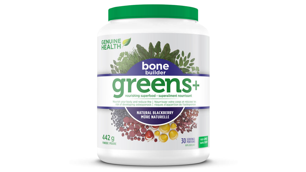
greens+™, bone builder™ et greens+bone builder™ ont tous des effets stimulants sur la formation osseuse dans les cellules ostéoblastiques humaines
(soumis par Rao LG le 30 septembre 2009)
Abstrait
INTRODUCTION
L'ostéoporose est une maladie squelettique caractérisée par une faible masse osseuse et une détérioration microarchitecturale du tissu osseux, entraînant une fragilité osseuse accrue et une augmentation conséquente du risque de fracture (1-3). La prévalence élevée de cette maladie et la morbidité associée à l'incidence des fractures coûtent 1,3 milliard de dollars par année au système de santé canadien (2). On estime que parmi la population canadienne actuelle de plus de 55 ans, une femme sur deux et un homme sur quatre souffrent d'ostéoporose (3). Étant donné que de nombreux nutriments ont été identifiés comme bénéfiques pour la santé osseuse (4), les bienfaits potentiels de l'intégration d'interventions nutritionnelles thérapeutiques aux traitements pharmaceutiques contemporains sont largement étayés par des données scientifiques. Un équilibre dynamique entre la formation et la perte osseuses est essentiel au maintien d'os sains et solides. L'alimentation est désormais reconnue comme un facteur important du mode de vie dans la gestion de la santé osseuse. Plusieurs études épidémiologiques ont démontré une association entre la consommation de fruits et légumes et la densité minérale osseuse (DMO) (5-7). Cela peut être attribué aux composés végétaux, nutritifs et non nutritifs, qui favorisent la formation osseuse ou réduisent la perte osseuse. Des compléments alimentaires, tels que greens+™, bone builder™ et greens+bone builder™, peuvent contribuer à un apport adéquat en nutriments, autrement manquants dans l'alimentation.
Le stress oxydatif a été impliqué dans la progression de l'ostéoporose. Un excès d'espèces réactives de l'oxygène, produites normalement par l'organisme lors de processus métaboliques, entraîne un stress oxydatif (8). Des études ont montré une corrélation inverse entre les biomarqueurs du stress oxydatif et la densité minérale osseuse (DMO) (9). Chez les patients présentant des fractures, les paramètres de stress oxydatif sont plus élevés que chez les témoins sains (10). Il a été démontré que les antioxydants capables de contrer cet effet, en neutralisant les espèces réactives de l'oxygène, jouent un rôle important dans la réduction du risque d'ostéoporose (11). L'accumulation d'espèces réactives de l'oxygène peut être contrecarrée par des antioxydants, principalement issus d'aliments tels que les fruits et légumes, ainsi que de boissons et de compléments alimentaires à base de plantes. Notre laboratoire étudie deux types d'antioxydants dont les effets bénéfiques sur la santé osseuse ont été démontrés : le lycopène liposoluble et les polyphénols hydrosolubles. Nous avons récemment démontré qu'un taux sérique élevé de lycopène est associé à une diminution de l'oxydation des protéines et de la résorption osseuse chez les femmes ménopausées (12), suggérant un potentiel bénéfique du lycopène dans la réduction du risque d'ostéoporose. Au niveau cellulaire, nous avons montré que le lycopène stimule la croissance et la différenciation des ostéoblastes (13,14) et inhibe la formation et la résorption des ostéoclastes (15). Le lycopène est l'un des composants de Bone Builder™. Les polyphénols ont également démontré des effets bénéfiques sur la santé osseuse (16). Le complément alimentaire greens+™ est un mélange de plusieurs produits à base de plantes et de plantes contenant une quantité importante de polyphénols qui agissent comme antioxydants et devraient donc pouvoir contrer le stress oxydatif. Notre laboratoire a démontré que les extraits polyphénoliques de greens+™ ont un effet stimulant sur la formation de nodules osseux minéralisés dans les cellules ostéoblastiques humaines, en fonction de la dose et du temps (17). Nous avons en outre montré que cet effet stimulant s'accompagne d'une diminution des espèces réactives de l'oxygène H2O2 (18), prouvant ainsi que greens+™ est capable de contrer le stress oxydatif dans les cellules ostéoblastiques humaines et peut donc être un bon candidat comme complément nutritionnel pour prévenir le risque d'ostéoporose.
Deux compléments nutritionnels supplémentaires ont depuis été formulés et pourraient s'avérer bénéfiques pour la santé osseuse. Il s'agit du Bone Builder™ et du Greens+Bone Builder™. Ce dernier est le produit Greens+™ original, complété par la formule Bone Builder contenant plusieurs composés, dont des vitamines, des minéraux et des antioxydants. Ces différents composants ont démontré séparément un effet bénéfique sur les os. Le calcium est le principal minéral formateur d'os. De nombreuses études ont montré que l'apport en calcium est positivement associé à une augmentation de la densité minérale osseuse (DMO) chez la majorité de la population mondiale (19-21). Cependant, des études ont montré que le calcium seul n'est pas suffisant, mais nécessite d'autres composants nutritionnels, pour augmenter la formation osseuse in vivo (22). Ainsi, les composants du Bone Builder™ autres que les trois calciums absorbables (citrate-malate, formiate et bisglycinate), notamment les vitamines D, C et B, le lycopène antioxydant, le magnésium, le sélénium, les oligo-éléments tels que le zinc, le cuivre et le manganèse, et le zinc. L'acide aminé essentiel, la L-lysine, le bore et le silicium se sont tous révélés importants, séparément, pour la formation osseuse, dont les mécanismes ne sont pas entièrement compris (revue dans 22). Cependant, leurs carences dans les modèles animaux entraînent des anomalies osseuses. Globalement, il ressort clairement de la littérature actuelle que d'autres nutriments, outre les recommandations standard en calcium et en vitamine D, pourraient être nécessaires à la santé osseuse. L'hypothèse de notre étude est qu'une combinaison des polyphénols antioxydants de greens+™ et des composants de Bone Builder aura, ensemble, un effet plus positif sur la formation osseuse que greens+™ ou Bone Builder™ seuls. L'objectif de notre étude était de comparer les effets séparés de l'extrait polyphénolique de greens+™ et du composant hydrosoluble de Bone Builder™ avec ceux de la combinaison des deux extraits (greens+bone builder) sur la formation osseuse par des ostéoblastes humains in vitro.
Matériels et méthodes
Les compléments alimentaires greens+™, bone builder™ et greens+ bone builder™ ont été fournis par Genuine Health Inc., Toronto, Canada. Tous les autres produits chimiques ont été achetés auprès de Sigma.
Préparation des échantillons
Préparation de l'extrait polyphénolique de greens+™ - greens+ ™ a été soumis à une extraction polyphénol-acétone, modifiée d'après Sun et al. (23). Brièvement, 1 g de greens+ a été agité pendant une heure avec de l'acétone à 80 % refroidie à l'aide d'un agitateur magnétique, puis filtré sur papier Whatman n° 2 dans un entonnoir Büchner sous vide. L'acétone-H2O a été évaporée à sec et le filtrat récupéré avec 50 ml d'H2O distillée (extrait de greens+). Une extraction parallèle a été réalisée sans greens+ pour servir de témoin. Les aliquotes ont été conservées à -20 °C. La teneur phénolique totale de l'extrait a été analysée par la méthode de Folin-Ciocalteu (24).
Préparation d'un extrait hydrosoluble de Bone Builder ™ – Les composants hydrosolubles du complément Bone Builder™ ont été dissous dans de l'eau bidistillée. Un g de Bone Builder pour 100 ml de solution a été préparé pour obtenir des solutions mères à 10 mg/ml par agitation sur un agitateur magnétique pendant 10 minutes, centrifugation pendant 10 minutes à 2500 tr/min, puis filtration sur seringues filtrantes de 0,2 µM dans des conditions stériles. L'extrait stérile a été réparti dans des tubes de 5 ml pour être congelé et conservé à -40 °C jusqu'à utilisation pour les expériences (Bone Builder ou extrait bb).
Culture cellulaire – Les cellules ostéoblastiques humaines, SaOS-2, ont été obtenues auprès de l'American Type Culture Collection (ATCC, Rockville, MD). Les cellules CD34+ ont été clonées dans notre laboratoire par dilution limite à partir d'une préparation commerciale de cellules hématopoïétiques. Les cellules ont été maintenues dans un incubateur à CO2 à 37 ºC avec une atmosphère de 5 % de CO2 et passées chaque semaine dans un flacon de 75 cm² dans du milieu F12 de Ham supplémenté avec 28 mM de tampon HEPES, pH 7,35, 1,1 mM de CaCl2, 2 mM de glutamine, 1 % de solution antibiotique-antimycosique et 10 % de sérum de veau fœtal (milieu) comme décrit précédemment (25). Lorsque les cellules devaient être utilisées pour les expériences, elles ont été étalées dans des boîtes de 12 puits dans un milieu supplémenté avec 10 nM de dexaméthasone et 50 µl/ml d'acide ascorbique. Le milieu a également été complété avec 10 mM de b-glycérophosphate au jour 8 et l'ajout a été répété à chaque changement de milieu jusqu'à la fin de la culture.
Traitement par extrait de greens+ (g+), solution hydrosoluble de reconstitution osseuse (bb) et combinaison d'extrait de greens+ et de solution de reconstitution osseuse (greens+bone builder ou g+bb) : des cellules SaOS-2 ou CD34+ ont été ensemencées à une densité de 1 x 10⁴ cellules/puits dans chaque puits d'une plaque à 12 puits. Différentes doses de g+, bb ou g+bb ont été ajoutées au jour 8 et à chaque changement de milieu par la suite. Les cellules ont été incubées à 37 °C et la culture a été arrêtée du jour 13 au jour 20 pour les dosages.
Détermination du nombre de cellules - Une modification du test au bleu de méthylène par Genty et al. (26) a été utilisée. À la fin de la période d'incubation, les cellules ont été lavées avec du PBS et fixées pendant la nuit avec du paraformaldéhyde à 4 %. Les cellules ont été rincées avec du PBS et colorées avec du colorant bleu de méthylène à 1 %, préparé dans un tampon borate 0,01 M, pH 7,5. L'excédent de colorant a été rincé toutes les heures avec le même tampon jusqu'à ce que la solution reste claire. La couleur bleue des noyaux a été éluée avec de l'éthanol-0,01 M HCl (1:1). L'échantillon a été dilué dans de l'éthanol-0,01 M HCl (1:1) et la densité optique de la couleur a été mesurée dans un spectrophotomètre Multiscan (Flow Laboratories, McLean, VA). Les résultats ont été calculés à partir d'une courbe standard du nombre de cellules connu en fonction de la lecture de la densité optique et corrigés du facteur de dilution.
Dosage de l'activité de la phosphatase alcaline (PAL) – Les cellules ont été rincées deux fois avec 50 mM de Tris(hydroxyméthyl)aminométhane (Tris), pH 7,4, récoltées par grattage et soniquées dans 0,5 ml de Tris contenant 0,05 % de Triton X-100. L'activité PAL a été déterminée selon la méthode de Lowry (27) en utilisant des dilutions appropriées de sonicats cellulaires, comme décrit précédemment (28). La libération de p-nitrophénol à partir de 10 nM de phosphate de p-nitrophényle dans un tampon contenant 1,0 mM de MgCl2.6H2O dans du 2-amino-2-méthyl-1-propanol, pH 10,3, a été réalisée à 35 °C. La réaction a été arrêtée après 5 minutes avec de la NaOH 0,5 N et l'absorbance a été mesurée à 405 nm à l'aide d'un lecteur de plaques Multiscan (Flow Laboratories, McLean, VA). La teneur en protéines des sonicats cellulaires a été déterminée à l'aide d'un réactif de coloration protéique commercial (Biorad). L'activité PAL a été exprimée en nanomoles par minute/mg de protéines.
Test de formation de nodules osseux minéralisés – Les cellules ont été lavées avec du PBS et fixées pendant la nuit avec du paraformaldéhyde à 4 %. Elles ont été colorées in situ avec du nitrate d'argent à 5 % et exposées aux UV pendant 1 heure. Les cellules ont ensuite été rincées à l'eau distillée et exposées à du thiosulfate de sodium à 5 % pendant 5 à 10 minutes pour éliminer le bruit de fond, selon la technique de von Kossa décrite précédemment (25). Les cellules ont ensuite été rincées et recouvertes de glycérol à 50 %. Les zones et le nombre de nodules minéralisés ont été quantifiés à l'aide d'un analyseur d'images (Fluorchem 8900, Alpha Innotech Corporation, Californie).
Analyse statistique – L'analyse de la croissance a été réalisée dans le cadre de deux expériences, chacune avec 3 répétitions. Les résultats ont été exprimés en moyenne + écart-type ou erreur-type de la moyenne, selon les expériences. Tous les graphiques ont été tracés avec Prism version 5 (Graphpad Software, San Diego, CA). Pour l'analyse statistique, une analyse de variance à un facteur (ANOVA) a été utilisée pour déterminer la significativité statistique des traitements et de l'évolution temporelle. Une valeur de p < 0,05 a été considérée comme statistiquement significative. Un test t ou des tests post-hoc incluant le test de Tukey ont été effectués.
RÉSULTATS
Publications
Les articles publiés et les présentations de résumés lors de réunions/symposiums locaux/internationaux liés au projet sont les suivants :
- Rao LG, B Balachandran, Rao AV 2009 L'extrait polyphénolique du complément nutritionnel greens+TM stimule la formation osseuse dans des cultures de cellules SaOS-2 de type ostéoblaste humain. Journal of Dietary Supplements 5(3) ; 264-282 ; 2008.
- Balachandran B, Rao V, Murray T, Rao LG 2004 Les polyphénols contenus dans l'extrait de la préparation à base de plantes greens+TM ont des effets sur la prolifération et la différenciation cellulaires
de la lignée cellulaire ostéoblastique humaine SaOS-2. Présenté lors de la 26e réunion annuelle de l'American Society for Bone and Mineral Research, du 1er au 5 octobre 2004, à Seattle.
Washington. (Journal de recherche sur les os et les minéraux 2004 19 (suppl): S403. -
Rao LG , Balachandran B, Rao AV. L'effet stimulant des polyphénols dans
l'extrait de préparation à base de plantes greens+TM sur le nodule osseux minéralisé
formation (MBNF) des cellules SaOS-2 est médiée par son effet inhibiteur sur la
espèces réactives de l'oxygène intracellulaires (iROS). Présenté lors de la 27e conférence annuelle
Réunion de la Société américaine de recherche sur les os et les minéraux, Nashville,
Tennessee, du 23 au 27 septembre 2005. -
Rao LG , Beca J. 2007 Le complément nutritionnel greens+ bone builderTM stimule
Formation osseuse dans des cultures cellulaires humaines SaOS-2 in vitro. Présenté au
Coalition canadienne des herbes, des épices et des produits de santé naturels : Tradition à
Technologie, Saskatoon, Saskatchewan, du 10 au 13 mai 2007. -
Le complément nutritionnel Rao LG greens+ bone builderTM stimule les os
formation dans des cultures cellulaires humaines SaOS-2 in vitro » présenté à Gelda
Scientific et Sunstar Inc, Mississauga, Ontario, 14 février 2007. -
Rao LG « Composants alimentaires naturels/compléments nutritionnels et santé osseuse »,
présenté au Collège canadien de médecine naturopathique, Grand rounds,
19 novembre 2007. -
Rao LG « Composants alimentaires naturels, compléments nutritionnels et mode de vie dans le
prévention de l'ostéoporose » présenté à l'Ostéoporose Support & Information
Groupe, Centre récréatif du village de Scarborough, Kingston Rd, Scarborough,
31 mars 2008. - Rao AV, Snyder DM, Mackinnon ES, Rao LG 2008 Oligo-éléments présents dans
les compléments nutritionnels stimulent la formation osseuse chez les ostéoblastes humains SaOS-2
cellules in vitro, présentation orale à la 5e Rencontre Internationale des Avancées
dans Antioxydants (oligo-éléments, vitamines et polyphénols) : moléculaire
mécanismes, aspects nutritionnels et cliniques qui se tiendra du 11 au 15 octobre 2008 à
Monastir-Sousse (Tunisie).
RÉSUMÉ DES ÉTUDES PRÉCÉDEMMENT PUBLIÉES SUR GREENS+™
Les résultats de ces études ont été publiés (17) et le manuscrit est annexé. Les principales conclusions de ces études sont que la prolifération cellulaire a été stimulée aux premiers stades (jours 2 à 4) et inhibée à un stade ultérieur (après le jour 7) du traitement par l'extrait polyphénolique de greens+ TM. L'inhibition du nombre de cellules a été suivie d'une stimulation initiale de la PAL par une concentration plus faible d'extrait de greens+ (jours 9 à 11). À un stade ultérieur du traitement (jour 13), greens+ TM a inhibé la PAL et stimulé la formation de nodules osseux minéralisés (jours 13 à 17) de manière dépendante du temps et de la dose. Les résultats étaient cohérents avec l'effet de greens+ TM sur la maturation plus rapide des ostéoprogéniteurs vers la progression vers un stade de formation osseuse par rapport aux cellules n'ayant reçu aucun traitement par greens+.
ÉTUDES SUR BONE BUILDER™
Deux types de cellules ostéoblastiques ont été utilisés pour cette étude. Nous avons d'abord utilisé des cellules SaOS-2, puis des cellules CD34+ afin de confirmer nos résultats sur une autre lignée cellulaire. Pour étudier l'effet du Bone Builder sur les cellules, nous avons utilisé son composant hydrosoluble (bb), comme décrit dans la méthode. Cette partie de l'étude s'est concentrée sur les effets du bb sur la formation osseuse. La formation osseuse par les ostéoblastes en culture peut être visualisée par coloration de von Kossa des nodules osseux minéralisés. Un exemple est présenté sur la microphotographie de cellules colorées par von Kossa et traitées avec des concentrations croissantes de bb de 0 à 1,0 mg/ml (Figure 1). On observe que l'intensité de la coloration, représentative de l'os formé par les ostéoblastes, augmente avec la concentration, de 0,5 mg/ml à 1,0 mg/ml. La surface des nodules colorés peut être quantifiée à l'aide d'un analyseur d'images. Dans les figures suivantes, les effets des traitements sont présentés sous forme de surface de nodules osseux minéralisés, quantifiée par analyse d'images. La figure 2 montre les effets, en fonction du temps et de la dose, de différentes concentrations de bb sur la surface de nodules osseux minéralisés dans les cellules SaOS-2. Au jour 17, les concentrations de traitement de 0,5 à 1,0 mg/ml étaient significativement différentes dans la zone de nodules minéralisés, par rapport aux témoins. Au jour 20, une plus grande gamme de concentrations de traitement était significativement différente des témoins (0,3 à 1,0 mg/ml). L'effet du traitement semble culminer à 0,8 mg/ml. Cependant, comme il n'y a pas de différences significatives entre la majorité des traitements allant de 0,7 à 1,0 mg/ml, il n'est pas possible d'être sûr de cette tendance. Français Il existe cependant un effet dose-dépendant significatif, après 17 et 20 jours de traitement (ANOVA à un facteur, p < 0,0001), avec une augmentation jusqu'à 500 fois au jour 17 et une augmentation de 1000 fois au jour 20 de la zone nodulaire par rapport au témoin. L'effet significatif au fil du temps (P < 0,05) a eu des changements drastiques similaires avec une augmentation de 3 fois de la zone nodulaire entre le jour 17 et le jour 20. L'effet stimulant de bb sur la formation osseuse minéralisée dans les cellules CD34+ de type ostéoblaste a été examiné et les résultats sont présentés dans la figure 3. Similaire à l'effet sur les cellules SaOS-2, bb a eu des effets dose-dépendants et dépendants du temps sur la formation osseuse minéralisée dans les cellules CD34+. Les résultats ont révélé que bien que le calcium soit aussi efficace que bb à toutes les concentrations aux jours 13 et 15 du traitement, il est devenu évident que le traitement par bb à une concentration de 1,0 mg/ml était plus efficace que le traitement au calcium après 17 jours. Ainsi, le calcium a stimulé la zone du nodule osseux minéralisé de 250 fois, tandis que la stimulation par bb à 1,0 mg/ml était supérieure à 1 000 fois. Cela pourrait indiquer que les autres composants du Bone Builder sont nécessaires à la stimulation de la formation osseuse.
ÉTUDES SUR GREENS+BONE BUILDER – COMPARAISON AVEC GREENS+™
OU CONSTRUCTEUR OSSEUX SEUL
Pour étudier l'effet de greens+ et de Bone Builder sur les cellules SaOS-2 ou Cd34 de type ostéoblaste, nous avons utilisé respectivement les polyphénols de greens+ (g+) extraits à l'acétone et l'extrait hydrosoluble de Bone Builder (bb). Pour étudier l'effet de Greens+ Bone Builder (g+bb), les extraits de g+ et de bb ont été ajoutés simultanément aux cellules. Nous avons étudié les effets des trois traitements sur la maturation des cellules vers le stade de formation osseuse, notamment sur la prolifération, l'activité de la phosphatase alcaline (ALP) et la formation de nodules osseux minéralisés. Comme le montre la figure 4, le traitement des cellules SaOS-2 par g+, bb et g+bb a entraîné une augmentation du nombre de cellules après le 13e jour. Après le 15e jour, aucune augmentation supplémentaire n'a été observée avec le traitement par g+ ou bb, et une inhibition a été observée uniquement avec le traitement par bb. La figure 5 illustre les effets des différents traitements sur l'ALP des cellules SaOS-2. Français Au jour 13, bb et g+bb avaient significativement diminué l'activité tandis qu'au jour 17, tous les traitements ont entraîné une PAL significativement plus faible par rapport au témoin. La signification statistique des valeurs de p est donnée dans le texte. L'effet de g+bb a été comparé à g+ seul en traitant les cellules avec différentes doses de g+ seul et différentes doses de g+ en présence de 0,5 mg/ml bb. Les effets stimulants dose-dépendants de différentes concentrations de g+ (ANOVA à un facteur, p < 0,001) et de différentes concentrations de g+ 0,5 mg/ml bb (ANOVA à un facteur, p < 0,0001) sur la formation de nodules osseux minéralisés dans les cellules SaOS-2 sont illustrés dans la Figure 6. À n'importe quelle concentration donnée de g+ utilisée, l'effet était plus élevé en présence de 0,5 mg/ml bb. L'effet de g+bb a ensuite été comparé à bb seul en traitant les cellules avec différentes doses de bb et différentes doses de bb en présence de 1,2 mg/ml g+. La figure 7 montre que différentes concentrations de bb sans (ANOVA à un facteur, p < 0,0005) ou avec 1,2 mg/ml g+ (ANOVA à un facteur, p < 0,001) ont des effets stimulants sur la formation de nodules osseux minéralisés dans SaOS-2, mais la présence de 1,2 mg/ml de greens+ avec bb à des concentrations de 0,5 et 0,8 mg/ml était plus efficace. À des concentrations de 1,0 mg/ml de bb, l'effet stimulant n'était pas significativement différent en présence ou en absence de 1,2 mg/ml g+.
Français Les effets de g+bb comparés à g+ seul ou à bb seul ont ensuite été étudiés dans les cellules Cd34+ de type ostéoblaste. La figure 8 illustre que l'ajout de 0,5 mg/ml de bb à différentes doses de g+ a multiplié l'effet par 10 à 0,8 mg/ml de g+ (p < 0,005), par 15 à 1,2 mg/ml de g+ (p < 0,005) et par 22 à 2,0 mg/ml de g+ (p < 0,001). Un effet dose-dépendant de g+bb (0,5 mg/ml de bb) s'est avéré significatif par ANOVA à un facteur (p < 0,005). Français La figure 9 illustre que l'ajout de 1,2 mg/ml de g+ à différentes doses de bb a multiplié l'effet par 5 à 0,5 mg/ml de bb (p < 0,05), par 15 à 0,7 mg/ml de bb (p < 0,05) et par 20 à 1,0 mg/ml de bb (p < 0,005). Un effet dose-dépendant de g+bb (0,5 mg/ml de bb) s'est avéré significatif par ANOVA à un facteur (p < 0,01). Ce lot particulier de cellules CD34+ présentait un très faible niveau de minéralisation, n'ayant que 250 pixels par rapport à la valeur de plus de 1500 pixels dans l'étude précédente (figure 3). Malgré cela, cependant, les effets synergétiques entre g+ et bb ont pu être observés.
DISCUSSION
Le résultat le plus important de cette étude est que, bien que greens+ et Bone Builder aient eu séparément des effets significatifs sur la stimulation de la formation de nodules osseux minéralisés dans les cellules SaOS-2 et CD34+, ces effets étaient significativement plus importants avec l'association des deux. Nous avons montré dans une étude précédente que greens+TM (17) avait un rôle stimulant sur la formation de nodules osseux minéralisés dans les cellules SaOS-2. Cependant, c'est la première fois que nous rapportons qu'une combinaison des deux produits était plus efficace que l'un ou l'autre. Les résultats ont été obtenus avec deux lignées cellulaires différentes, les cellules SaOS-2 et les cellules CD34+, démontrant que les effets sont reproductibles. Les trois traitements ont eu des effets stimulants sur la prolifération cellulaire plus tôt dans la culture. Les cellules ont été stimulées pour croître plus rapidement, de sorte qu'à des moments précoces, le nombre de cellules traitées était plus élevé que celui des témoins. Vers la fin des périodes de culture, les cellules témoins ont atteint des nombres statistiquement similaires à ceux des cellules traitées, suggérant que les cellules témoins pourraient avoir continué à proliférer alors que les cellules traitées ont maintenant cessé de croître et ont commencé à se différencier. Ces résultats concordent avec ceux concernant la phosphatase alcaline et la formation de nodules osseux minéralisés. Ils suggèrent que les cellules traitées ont proliféré plus tôt, ont mûri plus rapidement et ont pu ralentir leur croissance pour se différencier et se minéraliser plus tôt que les témoins. Il était important d'observer des diminutions marquées de la PAL lors des phases ultérieures de traitement par greens+, bone builder et greens+bone builder. La littérature scientifique montre que les cellules SaOS-2 se comportent phénotypiquement comme des ostéoblastes en passant par différents stades de prolifération suivis de différenciation en présence de dexaméthasone. Les stades de différenciation sont marqués par une augmentation des marqueurs ostéoblastiques, dont la PAL. Après la différenciation, la PAL diminue et la formation osseuse minéralisée a lieu. Ainsi, notre étude a démontré qu'à des phases ultérieures de 17 jours de culture, une réduction significative de la PAL a été observée pour les trois traitements par rapport au témoin. L'effet inhibiteur de greens+, bone builder et greens+bone builder sur la PAL, un marqueur de la formation osseuse, concorde avec leurs effets sur la maturation et la différenciation des ostéoblastes vers le stade de formation osseuse. Ainsi, la diminution de la PAL témoigne de la capacité des ostéoblastes à former des nodules osseux minéralisés. Ceci suggère que les cellules traitées se sont différenciées plus tôt en culture que les cellules témoins et qu'au moment de l'analyse, elles avaient terminé leur différenciation et commencé à former de l'os. Pour étudier les effets de greens+bone builder, nous avons dû combiner l'extrait polyphénolique de greens+ par extraction à l'acétone et l'extrait aqueux de bone builder, car les différents composants végétaux de greens+ et ceux de bone builder ne se prêtent pas à une seule méthode d'extraction. En effet, seul le composant hydrosoluble de bone builder a été étudié jusqu'à présent, et l'effet des composants liposolubles tels que la vitamine D et le lycopène devra faire l'objet de futures études. Par conséquent, l'effet stimulant du reconstructeur osseux rapporté ici aurait été bien plus important si du lycopène et de la vitamine D avaient été ajoutés. L'effet du composant hydrosoluble du reconstructeur osseux était supérieur à celui du calcium seul. Cela pourrait indiquer que les autres composants du reconstructeur osseux sont nécessaires à la stimulation de la formation osseuse.
Si nous avons constaté que greens+bone builder avait une plus grande capacité à stimuler la formation osseuse que greens+TM ou bone builderTM seuls, cela pourrait s'expliquer par le fait que le polyphénol antioxydant contenu dans greens+TM et les différents composants de bone builder, dont l'effet bénéfique sur la santé osseuse a été démontré individuellement, sont tous nécessaires à la formation osseuse par les ostéoblastes. Les mécanismes de cette synergie restent flous, mais nos résultats pourraient avoir des implications importantes dans la prise en charge de l'ostéoporose et suggérer que greens+bone builder peut être considéré comme un complément alternatif dans la prévention de l'ostéoporose, tant chez les hommes que chez les femmes ménopausées.
RÉFÉRENCES
1. Lane N. 2006 Épidémiologie, étiologie et diagnostic de l'ostéoporose. American Journal of Obstetrics and Gynecology 194 : S3-11.
2. Atkinson SA et Wendy WE. 2001 Nutrition clinique : 2. Le rôle de la nutrition dans la prévention et le traitement de l'ostéoporose chez l'adulte. Association médicale canadienne.
Journal 165 : 1511-1514.
3. Genuis SJ & Schwalfenberg GK. 2007 Choisir un os dans la gestion contemporaine de l'ostéoporose : stratégies nutritionnelles pour améliorer l'intégrité du squelette. Clinique
Nutrition 26: 193-207.
4. Palacious C. 2006 Le rôle des nutriments dans la santé osseuse, de A à Z. Critical Reviews in Food Science & Nutrition 46 : 621-628.
5. Zalloua PA, Hsu YH, Terwedow H, Zang T, Wu D, Tang G et al. 2007 Impact de la consommation de fruits de mer et de fruits sur la densité minérale osseuse. Maturitas 56 : 1-11.
6. Chen YM, Ho SC & Woo JLF 2006 Une consommation accrue de fruits et légumes est associée à une augmentation de la masse osseuse chez les femmes chinoises. British Journal of Nutrition 96 : 745-751.
7. Nieves JW 2005 Ostéoporose : le rôle des micronutriments. American Journal of Clinical Nutrition 81 : 1232S-1239S.
8. Sahnoun Z, Jamoussi K, Zeghal KM 1997 [Radicaux libres et antioxydants : physiologie humaine, pathologie et aspects thérapeutiques]. [Revue]. Thérapie 52 : 251-70.
9. Basu S, Michaelsson K, Olofsson H, Johansson S, Melhus H 2001 Association entre le stress oxydatif et la densité minérale osseuse. Biochem Biophys Res Commun
288:275-9.
10. Prasad G, Dhillon MS, Khullar M, Nagi ON 2003 Évaluation du stress oxydatif après fractures : une étude préliminaire. Acta Orthop Belg 69(6):546-551.
11. Maggio D, Barabani M, Pierandrei M, Polidori CM, Catani M, Mecocci P, Senin U, Pacifici R, Cherubini A 2003 Diminution marquée des antioxydants plasmatiques chez les personnes âgées
Femmes ostéoporotiques : résultats d'une étude transversale. J Clin Endocrinol Metab 88:1523-1527.
12. Rao LG, Mackinnon ES, Josse RG, Murray TM, Strauss A, Rao AV 2007 La consommation de lycopène diminue le stress oxydatif et les marqueurs de la résorption osseuse chez
femmes ménopausées. Osteoporos Int 18:109-115.
13. Kim L, Rao AV, Rao LG 2003 Lycopène II – Effet sur les ostéoblastes : Le caroténoïde lycopène stimule la prolifération cellulaire et l'activité de la phosphatase alcaline des cellules SaOS-2. J Med Food 6:79-86.
14. Park CK, Ishimi Y, Ohmura M, Yamaguchi M, Ikegami S 1997 La vitamine A et les caroténoïdes stimulent la différenciation des cellules ostéoblastiques de souris. J Nutr Sci et
Vitaminol 43:281-296.
15. Rao LG, Krishnadev N, Banasikowska K, Rao AV 2003 Lycopène I - Effet des ostéoclastes : Le lycopène inhibe la formation d'ostéoclastes stimulée par l'hormone basale et parathyroïdienne et la résorption minérale médiée par les espèces réactives de l'oxygène dans les cultures de moelle osseuse de rat. J Med Food 6:69-78.
16. Wu CH, Yang YC, WJ Y, Lu FH, Wu JH, Chang CJ. Preuves épidémiologiques d'une augmentation de la densité minérale osseuse chez les buveurs réguliers de thé. Arch Intern Med
2002;162:1001-1006.
17. Rao LG, Balachandran, B & Rao, AV 2008 L'extrait de polyphénols de légumes verts + supplément nutritionnel stimule la formation osseuse dans les cultures de type ostéoblaste humain
Cellules SaOS-2. Journal of Dietary Supplements 5 : 264-282.
18. Rao LG, Balachandran B, Rao AV 2009. L'effet stimulant des polyphénols contenus dans l'extrait de la préparation à base de plantes Greens+TM sur la formation de nodules osseux minéralisés (MBNF) des cellules SaOS-2 est médié par son effet inhibiteur sur les espèces réactives de l'oxygène intracellulaires (iROS). Présenté lors de la 27e réunion annuelle de l'American Society of Bone and Mineral Research, Nashville, Tennessee, du 23 septembre au 1er janvier 2019.
27, 2005.
19. Cashman KD 2006 Minéraux du lait (y compris les oligo-éléments) et santé osseuse. International Dairy Journal 16 : 1389-1398.
20. Welten DC Kemper HCG, Post GB & Van Staveren WA 1995 Une méta-analyse de l'effet de l'apport en calcium sur la masse osseuse chez les femmes jeunes et d'âge moyen et
hommes. Journal of Nutrition 125 : 2802-2814.
21. Rao LG, Khan T & Gluck G 2007 Le calcium provenant du minéral du lait LactoCalcium après digestion avec de la pepsine stimule la formation de nodules osseux minéralisés chez l'homme
Cellules SaOS-2 de type ostéoblaste in vitro et peuvent être rendues biodisponibles in vivo. Biosci., Biotechnol. Biochem. 71 : 336-342.
22. Graci S, DeMarco C, Rao LG 2006 La solution de renforcement osseux John Wiley & Sons, Canada, pp 39-46.
23. Sun J, Chu YF, Wu X, Liu RH. 2002 Activités antioxydantes et antiprolifératives des fruits courants. J Agric Food Chem 50:7449-7454.
24. Singleton VL, Orthofer R, Lamuela-Raventos RM. 1999 Analyse des phénols totaux et autres substrats d'oxydation et antioxydants au moyen du réactif de Folin-Ciocalteu. Méthodes. Enzymol 299:152-178.
25. Rao LG, Liu LJ, Murray TM, McDermott E, Zhang X. 2003 L'ajout intermittent d'œstrogènes, mais pas continu, stimule la différenciation et la formation osseuse dans les cellules SaOS-2. Biol Pharm Bull 26:936-945.
26. Genty C, Palle S, Vanelle L, Bourrin S, Alexandre C. 1994 Évaluation colorimétrique de cellules ostéoblastiques en culture (ROS 17/2,8). Biotech. Histochem 69:160-164.
27. Lowry. 1955 Microméthodes pour le dosage des enzymes. II. Procédures spécifiques : phosphatase alcaline. Méthodes Enzymol 4 : 371-372.
28. Rao LG, Sutherland MK, Reddy GS, Siu-Caldera ML, Uskokovic MR, TM M. 1996 Effets de la 1a,25-dihydroxy-16ène,23yne-vitamine D3 sur la fonction ostéoblastique des cellules SaOS-2 d'ostéosarcome humain - dépendance au stade de différenciation et modulation par le 17b-estradiol. Bone 19:621-627.

Figure 1. Une photomicrographie de cellules colorées par VonKossa a été prise au microscope après 17 jours de traitement avec des concentrations croissantes de préparation hydrosoluble de constructeur osseux, et les zones des nodules minéralisés colorés en noir ont été quantifiées à l'aide du système d'analyse d'images FluorChem 8900 (Alpha Innotech Corporation, Californie).

Figure 2. Effets dose-dépendants et temporels du reconstructeur osseux sur la surface des nodules osseux minéralisés dans les cellules SaOS-2. Indique une différence significative par rapport au véhicule : p < 0,0001 ; p < 0,0005 ; p < 0,005, #, p < 0,01 et ##, p < 0,05. Il existe un effet dose-dépendant significatif au jour 17 et au jour 20, selon l'ANOVA à un facteur ; p < 0,0001.

Figure 3. Effet d'une solution de reconstitution osseuse sur la formation osseuse des cellules CD34+, traitées à partir du 8e jour de culture pendant 13, 15 et 17 jours. Une analyse de variance unidirectionnelle a révélé un effet dose-dépendant du reconstitution osseuse à tous les temps (p < 0,0001). L'effet de 1,0 mg/ml de reconstitution osseuse est supérieur à celui du calcium seul après 17 jours de traitement, *p < 0,005.

Figure 4. Dosage de la prolifération cellulaire des cellules SaOS-2 traitées en continu à partir du 8e jour avec le milieu ©, l'extrait de greens+ (g+), le composant hydrosoluble du Bone Builder (bb) et une combinaison de g+ et bb (g+ plus bb). Le nombre de cellules a été déterminé par le dosage au bleu de méthylène, comme décrit dans le texte. Les cultures ont été arrêtées et dosées les 10e, 13e et 15e jours. Les résultats sont exprimés en moyenne ± erreur type de la moyenne (ETM) de trois réplicats de deux expériences distinctes (n = 6). * p < 0,05 par rapport au témoin.

Figure 5. Dosage de la phosphatase alcaline des cellules SaOS-2 traitées en continu à partir du jour
8 avec contrôle moyen ©, extrait de greens+ (g+), composant hydrosoluble du constructeur osseux
(bb) et une combinaison de g+ et bb (g+ plus bb). Les cultures ont été arrêtées et analysées.
les jours 13 et 17. Les résultats sont exprimés sous forme de moyenne ± SEM de trois réplicats chacun à partir de
Deux expériences distinctes (n = 5 ou 6). Une diminution significative de l'activité de la PAL a été observée.
comme suit : Jour 13 : c vs g, p = 0,54 (signification marginale) ; ***p<0,01 pour c vs bb et c
vs g+ plus bb et Jour 17 : # p< 0,05, pour c vs g+, ; ** p< 0,005, c vs bb, et *p<0,0005, pour c vs g+ plus bb.

Figure 6. Effet dose-dépendant de greens + (g+) avec et sans 0,5 mg/ml de constructeur osseux (bb) sur la zone de nodules osseux minéralisés dans les cellules ostéoblastiques SaOS-2. Des différences significatives ont été trouvées par rapport aux témoins respectifs comme suit : *p<0,0005, **p<0,005 ; ***p<0,05 ; #p<0,0001 ; ## p<0,001 ; ###p<0,01 ; Des différences significatives ont été trouvées lorsque le traitement avec g+ plus 0,5 mg/ml bb a été comparé au traitement avec g+ seul comme suit : a p<0,0001 ; b p<0,005 ; c p<0,01 ; d p<0,05. L'ANOVA à un facteur a montré un effet dose-dépendant de greens+ seul à p<0,001 et un effet dose-dépendant de greens+plus 0,5 mg/ml bb à p<0,0001.

Figure 7. Effets dose-dépendants du constructeur osseux (bb) avec et sans 1,2 mg de greens+ (g+) sur la zone de nodules osseux minéralisés dans les cellules ostéoblastiques SaOS-2. Des différences significatives ont été trouvées par rapport aux témoins respectifs comme suit : **p<0,005 ; ***p<0,05 ; ###p<0,01 ; Des différences significatives ont été trouvées lorsque le traitement avec bb plus 1,2 g+ a été comparé au traitement avec bb seul comme suit : b p<0,005 ; c p<0,01 ; d p<0,05. Des effets stimulants dose-dépendants de différentes concentrations de bb (ANOVA à un facteur, p<0,0005) et de différentes concentrations de bb plus 1,2 mg/ml g+ (ANOVA à un facteur, p<0,001) ont été observés.

Figure 8. Effet dose-dépendant de greens+ (g+) avec et sans 0,5 mg/ml de reconstructeur osseux (bb) sur la surface des nodules osseux minéralisés dans les cellules ostéoblastiques Cd34+. Des différences significatives ont été observées par rapport aux témoins respectifs, comme suit : Différence significative par rapport aux témoins respectifs : *p < 0,0005, **p < 0,005. Des différences significatives ont été observées lorsque le traitement par g+ plus 0,5 mg/ml de bb a été comparé au traitement par g+ seul, comme suit :
suit : bp< 0,005 ; a p< 0,001. L'ANOVA à un facteur a montré un effet dose-dépendant de greens+plus 0,5 mg/ml bb à p<0,0005.

Figure 9. Effets dose-dépendants du constructeur osseux (bb) avec et sans 1,2 mg de greens+ (g+) sur la surface des nodules osseux minéralisés dans les cellules ostéoblastiques CD34+. Des différences significatives ont été constatées par rapport aux témoins respectifs comme suit : **p<0,005 et *p<0,0005 ; Des différences significatives ont été constatées lorsque le traitement avec bb plus 1,2 g+ a été comparé au traitement avec bb seul comme suit : b p<0,005 ; et cp<0,05. Des effets stimulants dose-dépendants de différentes concentrations de bb plus 1,2 mg/ml g+ (ANOVA à un facteur, p<0,001) ont été observés.


